- Palsha Building, Oginga Odinga Street, Kisumu.
- +254 700 722 562 | +254 708 136 767
- bevixscientific@gmail.com

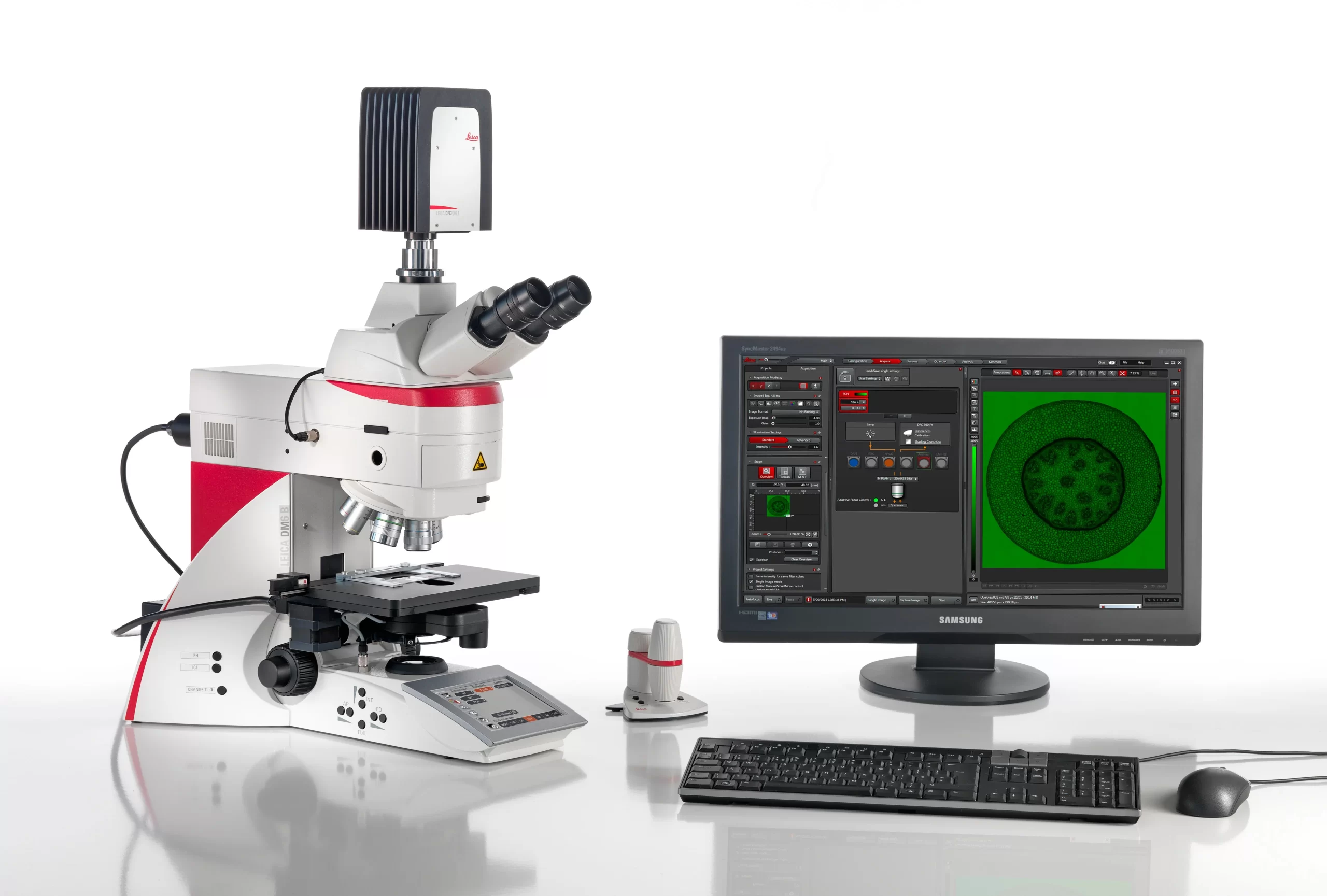
Fluorescence Microscope
A fluorescence microscope is a highly specialized optical imaging instrument that utilizes high-intensity light to excite fluorescent molecules (fluorophores) within a specimen. Unlike conventional microscopes that rely on transmitted or reflected light, it captures the longer-wavelength, lower-energy light emitted directly by the sample. This produces vibrant, high-contrast, luminescent images against a stark, dark background, enabling precise molecular targeting and submicrometer visualization.
Key Features & Core Components
- Integrated Filter Cube: Houses a precise combination of an excitation filter, a dichroic mirror (beam splitter), and an emission filter to cleanly isolate the weak emitted fluorescence from the bright excitation light.
- Versatile Light Sources: Utilizes high-power illumination options including mercury-vapor lamps, xenon arc lamps, and narrow-spectrum LEDs for widefield imaging, or advanced lasers for confocal setups.
- Epi-Fluorescence Design: Features an optimized optical path where both illumination and observation occur from above the sample, maximizing light collection through the objective lens.
- Multi-Channel Imaging: Includes manual or automated rotating turrets to easily swap filter cubes, allowing scientists to track multiple fluorescently tagged structures simultaneously within the same sample.
Primary Applications
- Live-Cell Imaging: Tracking dynamic cellular activities, biological signaling, and ion concentrations in real-time.
- Molecular Specificity: Localizing and mapping specific proteins, nucleic acids, and organelles tagged via immunofluorescence or genetic markers like GFP.
- Medical Diagnostics: Identifying cancer cells, tracking viral or bacterial infections, and diagnosing diseases by pinpointing abnormal structural biomarkers.
2000 in stock
Product successfully added to the Quote List
Add to cart